

The imaging research service encompasses a wide range of modern and classical methods that can capture spatial structures and properties at different scales – from single cells to entire landscapes. Typical fields of application on the landscape and field scales include, for example, earth surface surveying for the creation of 3D terrain models as well as erosion measurements using terrestrial or UAS (drone)-based laser scanners and photogrammetry, the determination of vegetation indices (e.g. NDVI) and plant physiological states by spectral imaging (VIS-NIR, hyperspectral), or the recording of surface temperatures using high-resolution thermography.
At the microscopic level, in addition to 2D imaging such as confocal, scanning electron and fluorescence microscopy, techniques such as holotomographic microscopy for imaging living cells and X-ray microtomography for detecting the internal structure of solids (e.g. porous media such as rocks or soils) are used.
X-ray photoelectron spectroscopy can also be used to spatially (2D) quantify the chemical compositions and bonding states of mineral and organic surfaces. Finally, methods of quantitative image analysis, which can be conducted (semi-)automated and AI-based, support the analysis of spatial structures and patterns in terms of their morphological and topological properties.
Devices, services and opportunities for cooperation
Showing results 21 - 28 out of 28
DJI Mini Pro 3
Burkhard, B. F. (Head)
Physical Geography and Landscape Ecology SectionFacility/Equipment: Research Instrumentation
Contact person(s) for booking: Benjamin Felix Burkhard
Atomic force microscope for biological applications NanoWizard® 4 AFM with Cellhesion (Bruker) and inverse AxioObserver3 fluorescence microscope (Zeiss), year of acquisition 2019
Lee-Thedieck, C. (Head)
Institute of Cell Biology and BiophysicsFacility/Equipment: Major Research Instrumentation
Contact person(s) for booking: Peter Schertl
Inverted confocal microscope with solid-state lasers 400, 488, and 512 nm, TE2000, Nikon, year of aqquisition 2005
Lee-Thedieck, C. (Head) & Ngezahayo, A. (Head)
Institute of Cell Biology and BiophysicsFacility/Equipment: Research Instrumentation
Contact person(s) for booking: Anaclet Ngezahayo
E-Mail(s): ngezahayocell.uni-hannover.de
LSM 980 inverted confocal microscope with incubation chamber and Airy Scan 2 super-resolution module (Zeiss), year of acquisition 2019
Lee-Thedieck, C. (Head)
Institute of Cell Biology and BiophysicsFacility/Equipment: Major Research Instrumentation
Contact person(s) for booking: Peter Schertl
E-Mail(s): schertlcell.uni-hannover.de
Inverted fluorescence microscope with monochromator and CCD camera (ORCA Hammatsu) Ti-E, Nikon, year of purchase 2013
Lee-Thedieck, C. (Head) & Ngezahayo, A. (Head)
Institute of Cell Biology and BiophysicsFacility/Equipment: Research Instrumentation
Contact person(s) for booking: Anaclet Ngezahayo
E-Mail(s): ngezahayocell.uni-hannover.de
Inverted fluorescence microscope with LED illumination and incubation chamber, AxioObserver 7, Zeiss, year of acquisition 2019
Lee-Thedieck, C. (Head)
Institute of Cell Biology and BiophysicsFacility/Equipment: Research Instrumentation
Contact person(s) for booking: Peter Schertl
E-Mail(s): schertlcell.uni-hannover.de
FE-TEM Feld-Emission Transmission Electron Microscope (JEOL JEM-2100F-UHR)
Feldhoff, A. (Head)
Institute of Physical Chemistry and ElectrochemistryFacility/Equipment: Research Instrumentation
Contact person(s) for booking: Armin Feldhoff
E-Mail(s): armin.feldhoffpci.uni-hannover.de
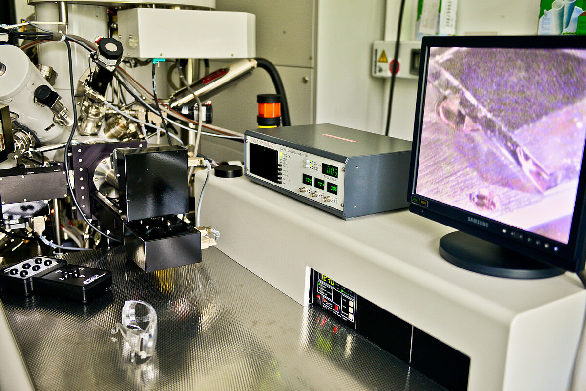
Scanning electron microscope JEOL JSM-7610F with Bruker EDX system
Holtz, F. (Head)
Mineralogy SectionFacility/Equipment: Research Instrumentation
Contact person(s) for booking: Renat Almeev
E-Mail(s): r.almeevmineralogie.uni-hannover.de


